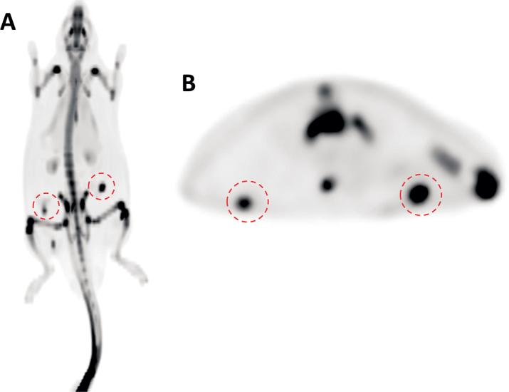
Figure 1.

PET images of the [18F]NaF uptake in grafts in the transverse (A) and coronal plane (B). Both of the sites (red circles) with implanted allogeneic bone (right side) and hydroxyapatite granules (left side) were easily distinguished in all animals.
