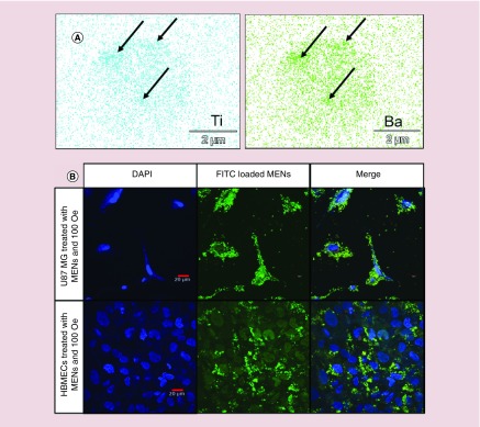
Figure 6. . Magnetoelectric nanoparticle uptake in malignant glioblastoma cells using a relatively weak d.c. magnetic field.
(A) EDS-SEM images of signature trace elements Ti and Ba in the cell lysate of U-87MG cells treated with MIA-GMO-MENs exposed to 100 Oe d.c. magnetic field. The arrows indicate where the MENs are most concentrated in the sample. (B) Confocal images showing the specific interaction of MENs with malignant glioblastoma cells. FITC-MENs are specifically associated with U-87MG cells. Uptake is not evident in nonmalignant HBMECs.
EDS: Electron-dispersive spectroscopy; FITC: Fluorescein isothiocyanate; GMO: Glycerol monooleate; HBMEC: Human brain microvascular endothelial cell; MEN: Magnetoelectric nanoparticle; SEM: Scanning electron microscopy.

