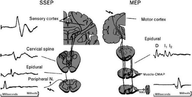
Figure 1.

Neuroanatomic pathways of somatosensory-evoked potential (SEEP) and motor-evoked potential (MEP). The SSEP is produced by stimulation of a peripheral nerve. The electrical signal travels through the dorsal columns of the spinal cord and crosses to the contralateral side after synapsing at the cervicomedullary junction. It ascends to another synapse in the thalamus and finally to the primary sensory cortex where the response is recorded. The MEP is produced by stimulation of the motor cortex in the brain. An electrical signal descends in the corticospinal tract to reach the anterior horn cells of the spinal cord. After synapsing, the signal travels along a peripheral nerve and across the neuromuscular junction to produce a muscle response. MEP signals can be recorded from the epidural space (D and I waves) or in the muscle (compound muscle action potential). Reprinted with permission from Sloan TB, Janik D, Jameson L. Multimodality monitoring of the central nervous system using motor-evoked potentials. Curr Opin Anesthesiol. 2008;21(5):560-564.
