
Figure 1. Four types of extravascular islands.
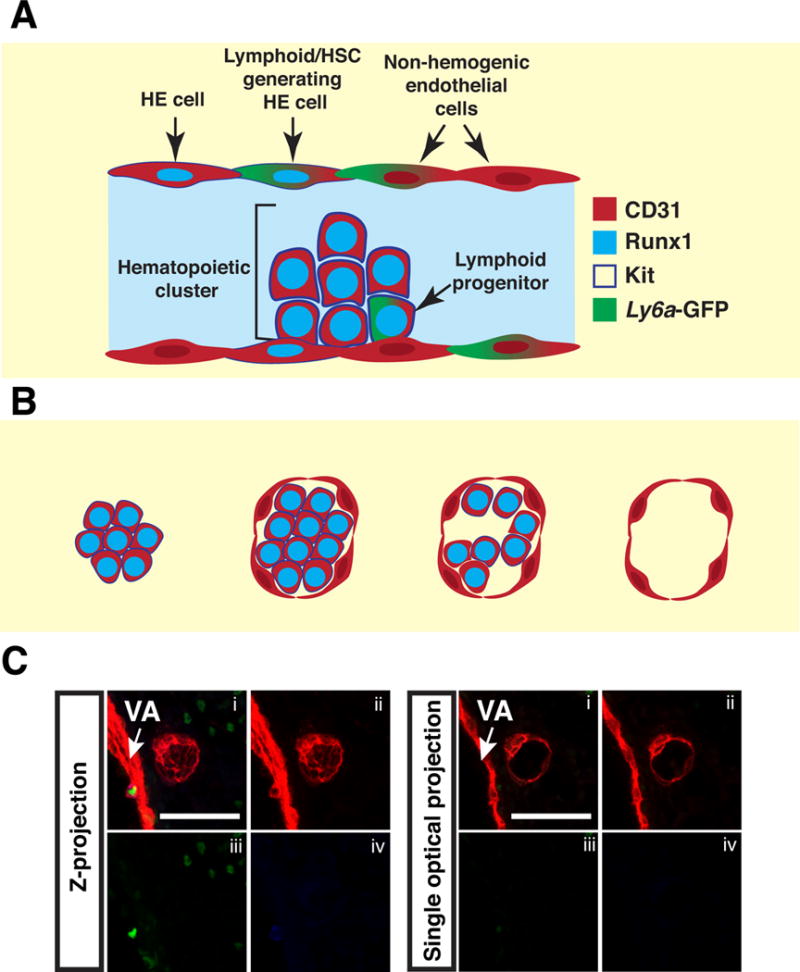
(A) Schematic illustrating the cell types identified by the combination of markers used in the analysis. (B) The four types of extravascular islands. Type I consists of tightly arranged hematopoietic cells with no endothelial component. Type II islands contain tightly arranged hematopoietic cells wrapped in a single layer of endothelial cells. Type III has loosely arranged hematopoietic cells wrapped in an endothelial layer. Type IV consists of a sphere of endothelial cells that are not associated with hematopoietic cells. (C) Z-projection of a type IV extravascular island that has been immunostained for CD31 (i,ii), Runx1 (i,iii) and Kit (i,iv). Panel on the right is a single optical projection illustrating the lack of Runx1+ Kit+ hematopoietic cells within a type IV extravascular island. Z-interval = 2μm and scale bar = 50μm.