
Figure 3. Extravascular islands do not contain Runx1+ hemogenic endothelial cells.
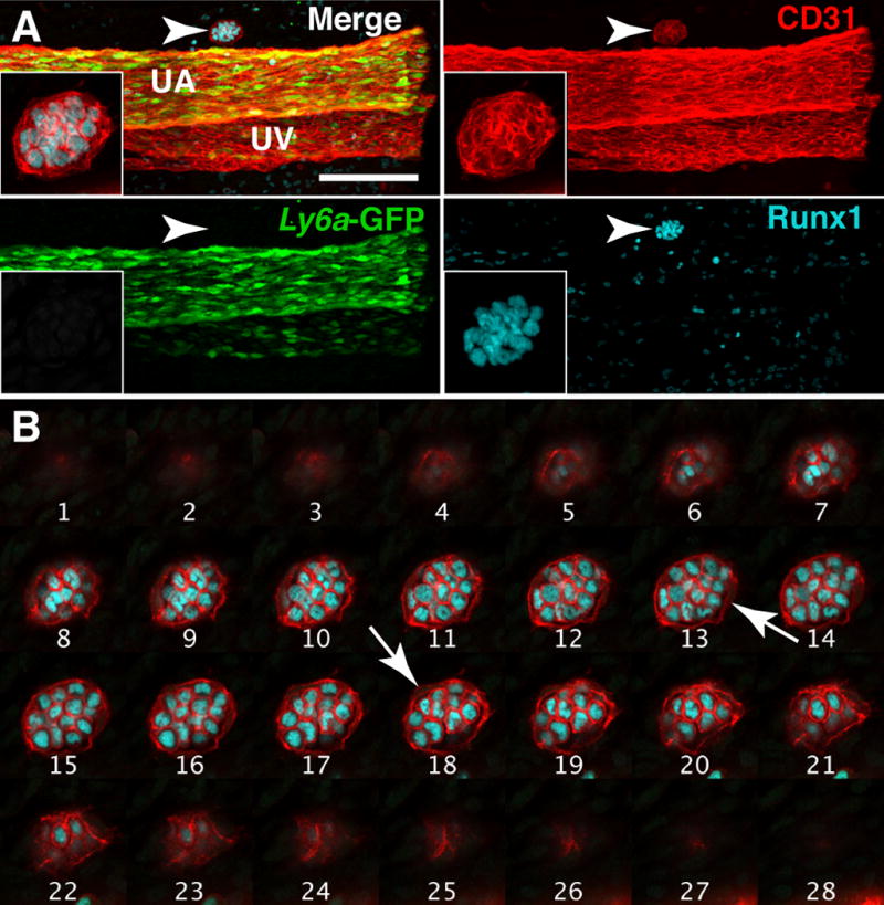
(A) Whole-mount Z-projection of the umbilical artery and vein of a 40sp Tg(Ly6a-GFP) embryo immunostained for CD31, Runx1 and GFP. Arrowhead indicates a Type II extravascular island on the cranial side of the umbilical artery. The extravascular island is located in between the embryo proper (which would be located to the left) and the chorion; inset is a magnified view of the extravascular island. Z-interval = 2μm, scale bar = 100μm; Z-interval of inset image = 1.17μm. UA = umbilical artery and UV = umbilical vein. (B) Montage of Z-slices through the extravascular island in (A). Each slice is 1.17μm apart. White arrows indicate endothelial cells.