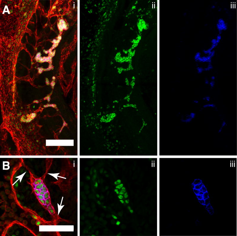
Figure 5. Hematopoietic clusters in the remodeling vitelline artery.

(A–B) Immunostaining for CD31 (i), Runx1 (i,ii) and Kit (i,iii). (A) Confocal Z-projection of the tributary vessels of the remodeling vitelline artery (VA) of a 34sp embryo. Z-interval = 5μm, scale bar = 100μm, DA = dorsal aorta. The embryo is oriented with its cranial end on the top and dorsal edge on the left. (B) Single optical projection of a hematopoietic cluster in the small tributary vessels of the vitelline artery of a 32sp embryo. Arrows indicate small endothelial projections that do not contain lumens. Scale bar = 50μm.
