
Figure 2. Tumor Growth Inhibition in C3−/− mice is mediated through CD4+ Lymphocytes.
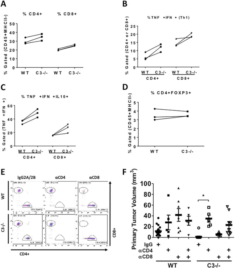
CMT-luc cells were injected into the lungs of WT or C3−/− mice 10 days prior to flow cytometric analysis of the immune populations using the sequential flow cytometry gating strategy shown in Supplemental Figure 2C. (A–D) Three individual experiments using separate isolations of T cells from 3 WT or C3−/− mice are shown. (A) The percentages of CD4+ or CD8+ cells are shown within the tumor-bearing lungs of WT and C3−/− mice. (B) CD4+ and CD8+ populations were analyzed for their ability to make TNFα and IFNγ after in vitro stimulation. (C) The TNFα+ and IFNγ+ CD4+ or CD8+ cells were gated for their ability to produce IL-10. (D) The percentages of CD4+ FOXP3+ (Tregs) are shown in both groups. (E) Representative flow cytometric plots of depletion efficacy measured with CD8 antibody clone (H35.17) and CD4 antibody (RM4-5) are shown (n = 2). The percentages of depleted T cells are shown in Supplementary Table 1. (F) The effects of in vivo depletion of CD4- and CD8- specific T cells on the primary CMT-luc volumes are shown (WT+IgG n = 16; WT+αCD4 n = 5; WT+αCD8 n = 7; WT+αCD4+αCD8 n = 5; C3−/−+IgG n = 14; C3−/−+αCD4 n = 5; C3−/−+αCD8 n = 5; C3−/−+αCD4+αCD8 n = 9). The depletion antibodies were administered starting a week before the tumor implantation, and CMT-luc tumors were harvested 3 weeks after implantation. *p <0.05. Error bars represent mean ± SEM