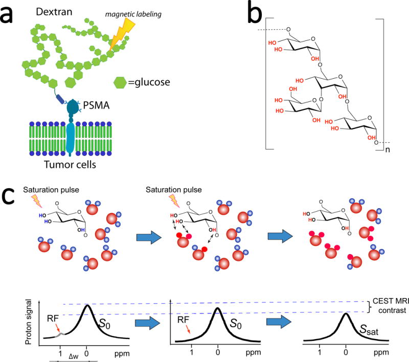
Figure 1. Development of dextran-based contrast agents for PSMA-receptor MR imaging.

a) Illustration of PSMA targeting and MRI detection of dextran-based agents that uses only RF-based magnetic labelling (signal saturation) without the need for metallic or radioactive labels. b) The chemical composition of dextran. c) Illustration of CEST MRI detection of natural dextran, which is achieved by the continuous transfer of saturated protons (red) from hydroxyl groups to surrounding water molecules, generating a reduction in the water signal (MRI contrast) proportional to the dextran concentration and rate of exchange.
