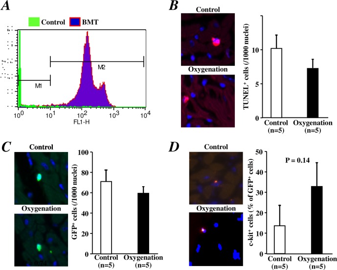
Fig 4. Histological analyses of cell apoptosis and the recruitment of bone marrow-derived (stem) cells 3 days after I/R injury of the heart.
A) A representative histogram of flow cytometry revealed that over 90% of circulating blood cells were replaced by GFP-positive cells at 6 weeks after post-transplantation with bone marrow cells from GFP-transgenic mice. B) Representative images (left) and quantitative data (right) of TUNEL-positive apoptotic cells in the LV anterior wall of the heart are shown. C) Representative images (left) and quantitative data (right) of bone marrow-derived GFP+ cells in the LV anterior wall of the heart are shown. D) Representative images (left) and quantitative data (right) of GFP+/c-kit+ stem cells in the LV anterior wall of the heart are shown.

