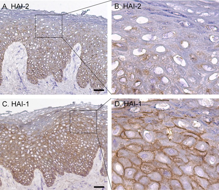
Fig 6. Expression pattern and subcellular localization of HAI-1 and HAI-2 in human skin.
Sections of human skin were immunostained with the HAI-2 mAb DC16 (A and B), the HAI-1 mAb M19 (C and D), and mouse IgG as negative control (data not shown). The cells were counterstained with hematoxylin. Representative examples of the staining observed are presented. Scale bar: 50 μm n>20.

