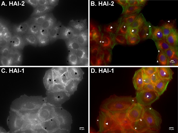
Fig 7. HAI-1 is primarily targeted to the intercellular contacts and HAI-2 largely remains inside HaCaT human keratinocytes.
The subcellular localizations of HAI-2 (A and B) and HAI-1 (C and D) in HaCaT human keratinocytes were analyzed by indirect immunofluorescent staining with the HAI-2-specific mAb DC16 and HAI-1-specific mAb M19, followed by Alexa 594-labelled anti-mouse IgG. The cells were also stained for F-actin using Alexa 488-labelled phalloidin (B and D, green) and nuclei using DAPI (B and D, blue), as counterstain. The staining is presented as black and white images (A and C) and merged false-color images (B and D). The staining of matriptase at different types of cell-cell contacts are as indicated. Scale bar: 10 μm.

