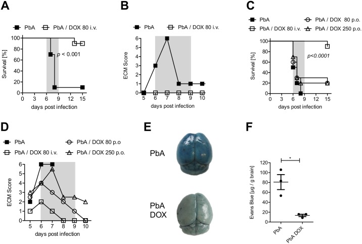
Fig 1. DOX administration reduced ECM development and stabilized the blood-brain barrier after PbA infection.
(A, B) C57BL/6 mice received 5*104 PbA-infected erythrocytes (PbA-iRBC) and indicated mice were injected i.v. with 80 mg/kg DOX dpi 4–6. All animals were then monitored for (A) survival and (B) ECM score (ECM phase dpi 6–9 marked in grey). (C, D) C57BL/6 mice received 5*104 iRBC and indicated groups of mice were treated orally with either 80 mg/kg/day or 250 mg/kg/day from dpi 4–6. Mice treated i.v. with 80 mg/kg/day DOX (dpi 4–7) served as reference treatment group. All mice were monitored for (C) survival and (D) ECM score (ECM phase dpi 6–9 marked in grey). Data sets are representative for 2–3 individual experiments with n = 10 mice/group. Survival data were analyzed with log-rank (Mantel-Cox) test. ECM scores are displayed as median. (E, F) Mice were infected with 5*104 PbA-iRBC ± 80 mg DOX/kg/day. The integrity of the BBB was analyzed with an Evans Blue assay on dpi 6. All mice were injected i.v. with 2% Evans Blue dye and one hour later, extravasation of the dye into the brain was determined. (E) Photo documentation of the discoloration and (F) quantification of dye extravasation into the brain by measuring the absorbance of brain tissue at 620nm. Data show representative results of 1 from 2 independent experiments. *p<0.05, t-test on data after analysis of normal distribution.

