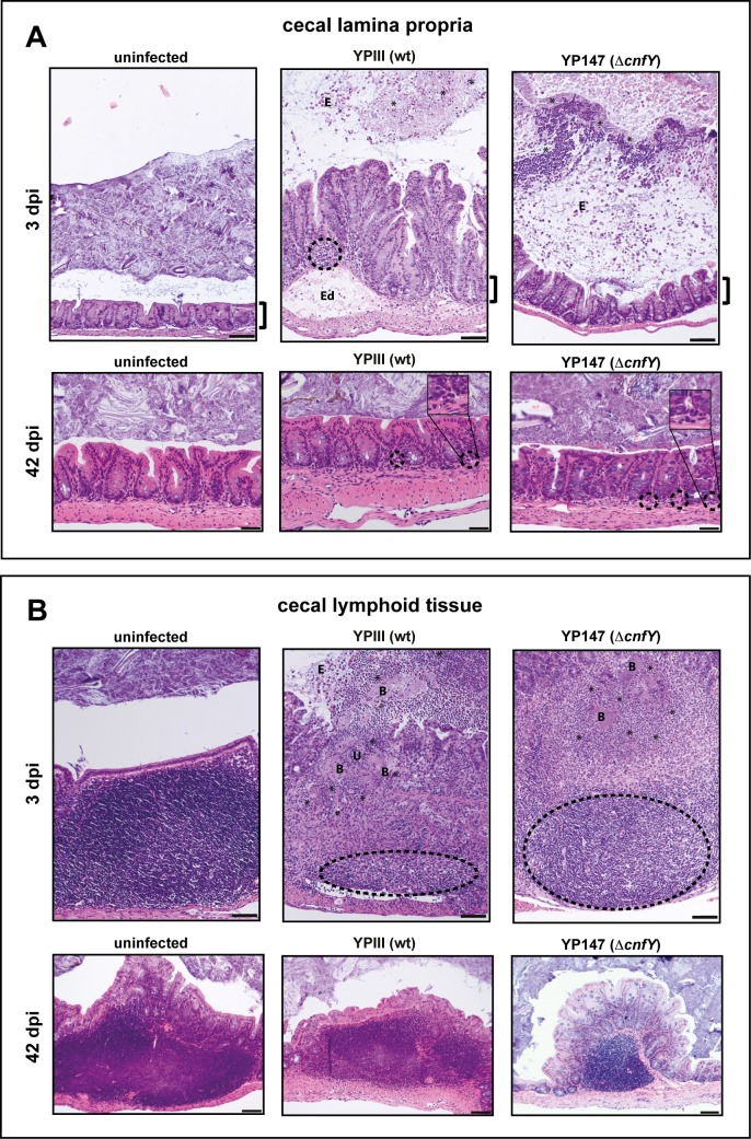
Fig 2. Tissue alterations during acute and persistent infection of the cecum.
H&E stained sections of the cecal lamina propria (A) and the cecal lymphoid tissue (B) of BALB/c mice at 3 or 42 dpi with about 105−106 CFUs of YPIII or YP147(ΔcnfY)/g tissue, or uninfected mice. Cecal lamina propria (3 dpi); YPIII: focal invasion of lymphocytes into the lamina propria (dashed halo) and edema formation (Ed). YP147(ΔcnfY): diffuse distributed granulocytes. E: epithelial cells. Cecal lamina propria (42 dpi); YPIII and YP147(ΔcnfY): isolated granulocytes at the basal lamina propria (dashed halo). Cecal lymphoid tissue (3 dpi); YPIII: massive necrosis, destroyed follicles (dashed halo), ulcus formation (U) and bacterial microcolonies (B) surrounded by invaded granulocytes (black asterisks). E: epithelial cells. YP147(ΔcnfY): necrotic parts and reduced lymphocytes in follicle (dashed halo). Infiltrating granulocytes surround bacterial microcolonies (black asterisks). Pictures show representatives of multiple fields of sections from groups of 3–5 mice. The brackets illustrate the length of the microvilli of the uninfected mice during the acute infection phase. Bar: (A) 50 μm, lower panel, 100 μm upper panel, (B) 100 μm.

