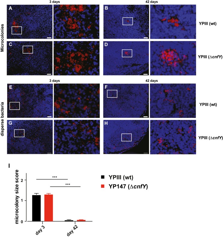
Fig 3. Colonization patterns of Y. pseudotuberculosis during acute and persistent infection.
Microscopic detection of mRuby2-expressing Yersinia in cecal lymphoid tissue of infected BALB/c mice at 3 and 42 dpi with approximately 105−106 bacteria/g tissue. Blue: DAPI-stained host cell nuclei; red: mRuby2-expressing bacteria. Bar: 50 μm. Microcolonies of YPIII (mRuby2) 3 dpi (A) or 42 dpi (B), YP147(ΔcnfY) (mRuby2) 3 dpi (C), or 42 dpi (D). Disperse bacteria of YPIII (mRuby2) 3 dpi (E) or 42 dpi (F), YP147(ΔcnfY) (mRuby2) 3 dpi (G) or 42 dpi (H). Representatives of multiple sections from groups of 3 mice are shown. (I) Scoring of the Yersinia colonization pattern of multiple microscopic sections of YPIII (mRuby2)- or YP147(ΔcnfY) (mRuby2)-infected mice (3 mice/group) at 3 or 42 dpi (Supplemental Information). Number of scored microscopy fields for microcolonies 3 dpi: YPIII n = 141, YP147(ΔcnfY) n = 237; 42 dpi: YPIII n = 166, YP147(ΔcnfY) n = 167. Data show the mean of scores ±SEM. Statistical analysis was performed using multiple t-tests, Holm-Šídák correction; P-values: * <0.05, ** <0.01, *** <0.001.

