. 2014 Nov 7;6(2):1206–1211. doi: 10.1039/c4sc03375b

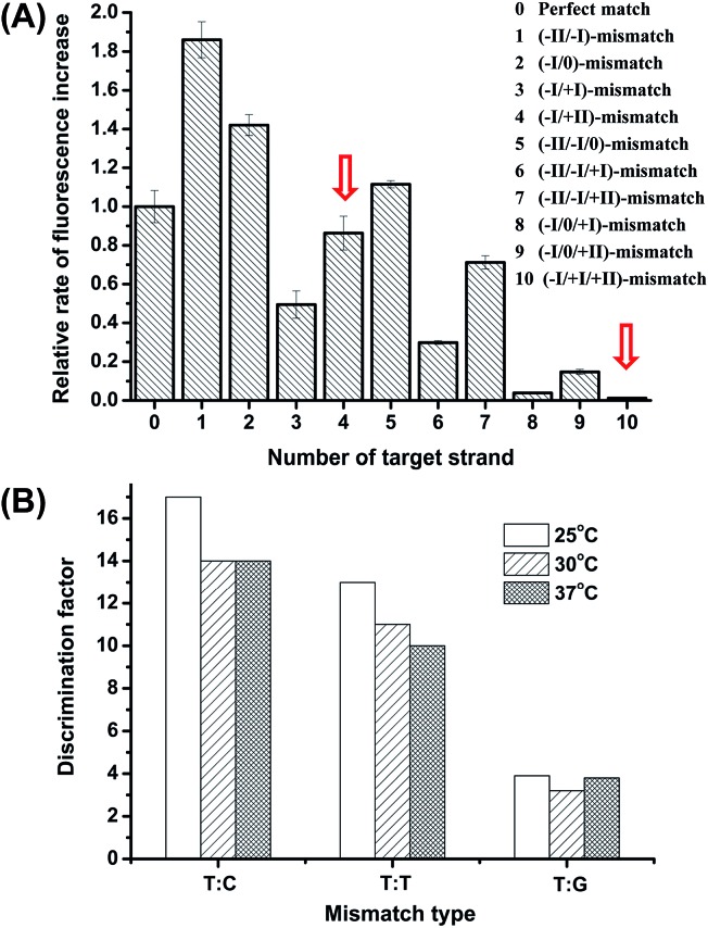
Fig. 2. (A) Comparison of the rates of the fluorescence increase for the probe/target hybrid containing double or triple mismatches at different positions in comparison with the perfectly matched duplex. The Roman numerals in the brackets indicate the positions of the mismatch bases in the target strands when hybridized to probe P-3FAM-4C (the sequences is 5′PO4-TCT(-FAM)CCACAGACACATACTCCA-BHQ1). The two red arrows denote the data observed for the probe/target hybrids containing the (–I/+II)-double-mismatch and the (–I/+I/+II)-triple-mismatch, respectively. (B) Comparison of the DFs between the (–I/+II)-double-mismatch and the (–I/+I/+II)-triple-mismatch at different temperatures. P-3FAM-4T(5′PO4-TCT(-FAM)TCACAGACACATACTCCA-BHQ1) was used for the test. The two purposefully inserted mismatched bases are underlined and the base opposite the SNP site is indicated in bold italics.

Fig. 2