
Figure 5. Cell print viability assay.
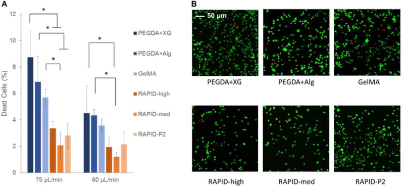
A. Quantification of LIVE/DEAD staining of 3T3 cells printed through a 32-gauge nozzle at average speeds of 75 and 60 μL/min. All bio-inks show less than 10% cell death during printing, while RAPID inks show less than 4% cell death. B. Representative LIVE/DEAD confocal images of 3T3 cells printed at a flow rate of 75 μL/min immediately after print extrusion and before any curing steps.