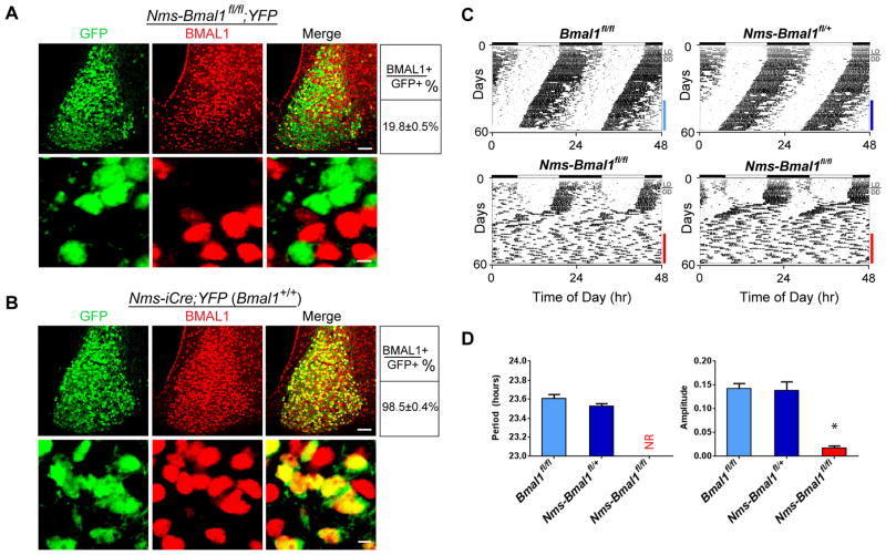
Figure 3. Loss of Bmal1 in Nms Neurons Abolishes Behavioral Circadian Rhythms.
(A) Double staining of YFP (green) and BMAL1 (red) on SCN sections of Nms-Bmal1fl/fl;YFP mice using anti-GFP and anti-BMAL1 antibodies, respectively. The scale bars are 50 μm (top) and 5 μm (bottom). Quantitative data are shown at the right.
(B) Double staining of YFP (green) and BMAL1 protein (red) on SCN sections of Nms-iCre;YFP mice using anti-GFP and anti-BMAL1 antibodies, respectively.
(C) Representative actograms of Nms-Bmal1fl/fl (bottom) along with Bmal1fl/fl (top-left) and Nms-Bmal1fl/+ mice (top-right). Colored bars represent the days of analyses presented in (D).
(D) Quantification of circadian activity in Nms-Bmal1fl/fl, Bma11fl/fl, Nms-Bmal1fl/+ mice. Nms-Bmal1fl/fl exhibited no coherent rhythms (NR) and a low average amplitude in the circadian range compared to Bmal1fl/fl and Nms-Bmal1fl/+ control mice (one-way ANOVA, *P<0.05 by Tukey’s post-hoc test). Values are mean ± SEM (Bmal1fl/fl, n=7; Nms-Bmal1fl/+, n=6; Nms-Bmal1fl/fl, n=14).

