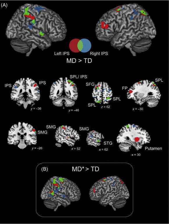
FIG. 4.

Parietal hyperconnectivity in children with MD. Brain areas that showed greater IPS connectivity in children with MD compared to typically developing (TD) children. (A) Children with MD showed hyperconnectivity between bilateral IPS and multiple dorsal frontal and parietal cortical regions, between bilateral IPS and right hemisphere SMG and STG, and between left IPS and right putamen. (B) Results were almost identical when examining a subset of 14 MD children who scored at or below 85 on the numerical operations subtest of the WIAT-II (MD*). Greater connectivity for MD>TD in red (dark gray in the print version) (left IPS), blue (gray in the print version) (right IPS), and green (light gray in the print version) (both left and right IPS). Coordinates are in MNI space. FP, frontal pole; IPS, intraparietal sulcus; SFG, superior frontal gyrus; SMG, supramarginal gyrus; SPL, superior parietal lobe; STG, superior temporal gyrus.
Adapted from Jolles, D., Ashkenazi, S., Kochalka, J., Evans, T., Richardson, J., Rosenberg-lee, M., Zhao, H., Supekar, K., Chen, T., Menon, V., 2016. Parietal hyper-connectivity, aberrant brain organization, and circuitbased biomarkers in children with mathematical disabilities. Dev. Sci.
