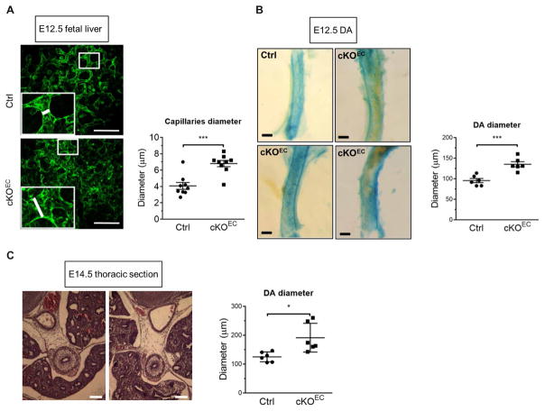
Fig. 2. Depletion of Drosha in the endothelium in mice results in embryonic developmental vascular abnormalities similar to HHT.
(A) Representative images of a whole-mount liver from control (Ctrl) or Drosha cKOEC embryos at embryonic day (E) 12.5 stained with CD31 are shown. Quantitative analysis of the diameter of fetal liver capillaries is shown as means ± SEM. Scale bar, 100 μm. n = 4 Ctrl embryos and 3 cKOEC embryos. (B) β-Galactosidase staining was performed to visualize the dorsal aorta (DA) isolated from Ctrl or cKOEC embryo at E12.5. Quantitative analysis of the DA diameter is shown as means ± SEM. ***P < 0.001, significant by two-tailed unpaired Student’s t test. Scale bar, 50 μm. n = 4 Ctrl embryos and 4 cKOEC embryos. (C) Representative hematoxylin and eosin (H&E) stain images of a 7-μm transverse thoracic section of Ctrl or cKOEC embryos at E14.5. Scale bar, 100 μm. The diameter of DA was quantified by ImageJ and presented as means ± SEM. n = 3 Ctrl embryos and 3 cKOEC embryos. *P < 0.05, significant by two-tailed unpaired Student’s t test. Hemorrhagic telangiectasia, HHT.

