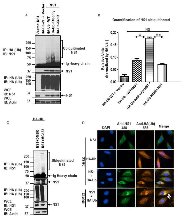
Figure 3. DENV-NS1 is ubiquitinated with K48 poly-Ub chains in cells and co-localizes with ubiquitin.
A–C) HEK293T cells were transfected with NS1 and HA-Ub-WT, HA-Ub-K48only, and HA-Ub-K48R. Twenty-four hours post-transfection, cells were treated with either DMSO (control, panel A) or 20 μM MG132 for an additional 6 h (panel C). Cells were harvested and whole cell extracts (WCE) were used for HA-Ub pull-down using HA-beads. B) Quantification of ubiquitinated NS1 was performed using ImageJ software. To normalize the efficiency of HA-Ub pulldown, the densitometry values obtained for the full smear of ubiquitinated NS1 (top blot, IP: HA, IB: NS1) were normalized by the levels of immunoprecipitated HA-Ub (IP: HA, IB: HA). The quantification was performed for two independent experiments and SEM is shown. Statistical significance was determined in Graphpad software. *, P < 0.05; **, P < 0.01; ***, P < 0.001; NS, not significant. D) Localization of NS1 with ubiquitin. HeLa cells were transfected with NS1 and HA –Ub for 24 h, followed by treatment with DMSO or MG132 for an additional 6 h. The cells were then fixed and stained with the indicated antibodies. The cells were visualized in a fluorescent microscope. The results shown are representative of 2 independent experiments.

