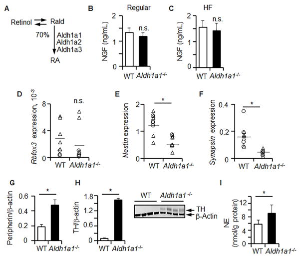
Figure 1. Aldh1a1 deficiency induces innervation in WAT in vivo without an increase in neural precursors.
(A) Schematic of the main processes leading to production of retinoic acid (RA) in WAT. ALDH1A1 produces 70% of RA which activates RAR in adipocytes (100%) [20]. NGF plasma levels were examined by ELISA in WT and Aldh1a1−/− mice fed regular chow (B) or HF diet (C), n=4 per group randomly selected from Study 1 and 2. Relative expressions of Rbfox3 (D), Nestin (E), and Synopsin (F) were measured in iAb WAT from WT and Aldh1a1−/− males (triangle, n=5) and females (circle, n=5) by TaqMan RT-PCR (HF, Study 1). Expressions were normalized to Tata box protein (TBP). The levels of peripherin (G) and tyrosine hydroxylase (TH) (H) were analyzed by Western blot from randomly selected tissues of WT (n=4) and Aldh1a1−/− mice (n=4) (HF, Study 1). Protein expression was normalized by levels of housekeeping genes (β-actin). The insert shows representative Western blot. (I) Norepinephrine (NE) level from iAb fat pat homogenates in WT (n=6) and Aldh1a1−/− mice (n=5) was measured using ELISA (HF diet, Study 1). An asterisk indicates a significant difference between WT and Aldh1a1−/− groups (p<0.05). n.s.: not significant, Mann-Whitney U test.

