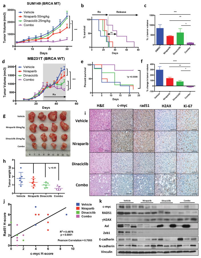
Figure 5.
Dinaciclib + Niraparib treatment in vivo (a–c) SUM149 cells were injected into immunocompromised mice and allowed to grow to ~200mm3, xenografts were treated with either Vehicle, Dinaciclib 25mg/kg 3 times weekly, Niraparib 5mg/kg 5 times weekly or Dinaciclib + Niraparib (Combo) therapy for 4 weeks (a) Tumor volume measurements and (b) Kaplan Meyer survival analysis of mice treated with indicated drug treatment arms for 4 weeks (28 days), followed by release without treatment until mice reached maximum allowed tumor size (c) Percent change in tumor volume normalized by day 1 of treatment. *P < 0.05, **P < 0.01, ***P < 0.001 two-way ANOVA with Sidak post-test correcting for multiple comparison (d–j) MB231T cells were injected into nude mice and cells were allowed to grow to >500mm3 before treatment was initiated. Mice were treated with Vehicle, Dinaciclib 25mg/kg 3 times weekly, Niraparib 5mg/kg 5 times weekly or Dinaciclib + Niraparib (Combo) therapy for 3 weeks. Upon completion tumors were extracted and analyzed by IHC and RPPA (d) Tumor volume measurements and (e) Kaplan Meyer survival analysis of mice treated with indicated drug arms for 3 weeks (f) Percent change in Tumor volume normalized by Day 1 of treatment. *P < 0.05, **P < 0.01, ***P < 0.001 two-way ANOVA with Sidak post-test correcting for multiple comparison (g–h) Tumors from mice treated with each of the treatment arms were extracted and weighed and subjected to (i) Immunohistochemical analysis of treated tumors for H&E, Ki67, c-myc, RAD51, and γHAX resulting in a (j) comparison the H-score of c-myc with RAD51 expression in MB231T treated xenografts (k) immunoblot analysis for c-myc, RAD51, γH2AX, Zeb1, Axl, E-cadherin, N-cadherin and Actin.

