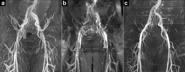
Figure 8.

Ungated free-breathing QISS FISS MRA of the pelvis. Comparison of (a) ungated radial QISS FISS MRA (α=70°, n=3, RF spoiling) and (b) ungated radial QISS bSSFP MRA (α=70°; 1.6-fold more views acquired in each shot to match the readout duration of QISS FISS) with respect to a reference exam of (c) cardiac-gated breath-hold Cartesian QISS bSSFP MRA (α=80°). Acquisitions in (a) and (b) were acquired without breath-holding, while the upper-most slices in (c) were acquired with breath-holding. Whereas ungated radial QISS bSSFP MRA shows severe artifact, ungated radial QISS FISS MRA displays the pelvic arteries, and agrees with the cardiac-gated breath-hold Cartesian QISS bSSFP MRA.
