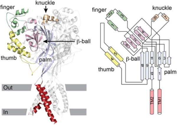
Figure 1.

ASIC1 structure. Left, cartoon of ASIC1 trimer. Domains of one subunit highlighted. Approximate location of the membrane is indicated. Right, schematic of an ASIC1 subunit. Cylinders represent helices, arrows represent β strands. The peripheral finger, knuckle and thumb domains are contiguous α helices arising from the central β-ball or palm domains. Modified from (1).
