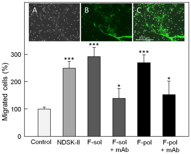
Fig. 7.

Effect of soluble fibrin and endothelium-anchored fibrin polymers on transendothelial migration of leukocytes. HUVECs were grown to confluence on gelatin-coated cell culture inserts, Calcein AM-labeled differentiated HL-60 cells were added to the upper chambers on top of the HUVEC monolayers in the presence of PBS or 1.5 μM NDSK-II (controls), or 1.5 μM soluble fibrin without (F-sol) and with 0.5 μM mAb 1H10 (F-sol + mAb). Fibrin polymers (F-pol) were deposited (anchored) on the HUVEC monolayers using the procedure described in Materials and methods and tested without or with 0.5 μM mAb 1H10 (F-pol + mAb). The cells were allowed to migrate into the lower chambers containing chemoattractant fMLP for 4 h at 37°C, collected, and quantified as described in Materials and methods. The results are expressed as percentage of HL-60 cells migrated in the presence of PBS (control). The graph shows combined data from 2 independent experiments; error bars denote means ± SD (n = 6). *P < 0.05; ***P < 0.001. The presence of endothelium-anchored fibrin polymers on the HUVEC monolayers was confirmed by fluorescence microscopy as shown in the insets. Inset A shows representative image of membrane insert with bright and dark dots representing membrane pores and differentiated HL-60 cells adherent to the endothelial surface, respectively. Inset B, the same field as in inset A showing endothelium-anchored fibrin polymers labeled with Alexa-488 that appear on the surface as a fiber network colored in green; Calcein AM-labeled differentiated HL-60 cells appear as green dots. Inset C shows merged insets A and B. All images were taken at ×10 magnification using an EVOS FL Auto microscope. Scale bar: 200 μm.
