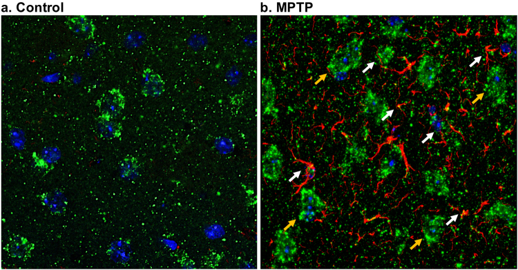
Figure 7.
Co-localization of glutamate and GFAP. The confocal microscopy of the brain slices were performed in a 63 × zoomed region in the striatum of the control (a) and MPTP treated mouse (b). The figure shows the cell bodies with nuclear stain DAPI (blue) having glutamate (green) and GFAP (red) immunostaining. Neuronal nuclei are bigger and are found to be co-localized with neuronal glutamate (yellow arrows), whereas, astrocytic glutamate appeared to co-localize with GFAP staining. Areas marked with white arrows show the co-localization of glutamate and GFAP in the reactive glia.

