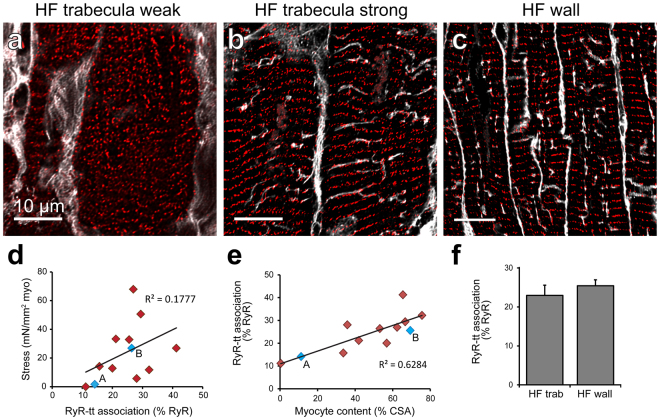
Figure 4.
Structural correlates with contractile function in human cardiac trabeculae. Deconvolved confocal images show t-tubule (WGA; grey) and RyR (red) dual labelling in cardiomyocytes from trabeculae exhibiting either (a) weak or (b) strong contractile function, as well as (c) ventricular myocardium from the human failing heart. (d) Stress normalised to MCSA forms a weak, positive relationship with the percentage of RyR clusters associated with t-tubule labelling (p = 0.167). (e) A strong positive correlation is observed between increasing myocyte content as a percentage of total CSA and the degree of association between RyR clusters and t-tubules (p = 0.0014). Blue data-points labelled ‘A’ and ‘B’ indicate values from corresponding images. (f) Analysis of RyR-t-tubule association was performed in the sample groups. RyR-t-tubule association analysis: Trabeculae: n = 22 images, 14 trabeculae, 5 hearts; failing wall n = 28 images, 5 hearts; Data displayed as mean ± SEM.

