Figure 5.
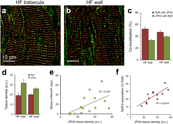
RyR co-localisation with JPH2 is altered in human HF. Deconvolved confocal micrographs showing RyR (red) and JPH2 (green) dual labelling in (a) a trabecula and (b) ventricle wall sample from failing hearts. (c) Co-localisation analysis reveals the extent of RyR co-localised with JPH2 and JPH2 co-localised with RyR in the trabeculae and ventricle wall from failing human hearts. (d) Tissue labelling density revealed no differences for RyR or JPH2. (e) A positive relationship is observed between JPH2 tissue density and peak stress development (normalised to MCSA) in trabeculae from the failing human heart (p = 0.041). (f) JPH2 tissue labelling density forms a strong, positive relationship with the percentage of RyR clusters associated with t-tubules (tt) in failing human trabeculae (p = 0.0014). Co-localisation analysis: Trabeculae: n = 21 images, 14 trabeculae, 5 hearts; failing wall n = 22 images, 5 hearts. Density analysis: Trabeculae: n = 14 images, 14 trabeculae, 5 hearts; failing wall n = 14 images, 5 hearts. Data displayed as mean ± SEM.

