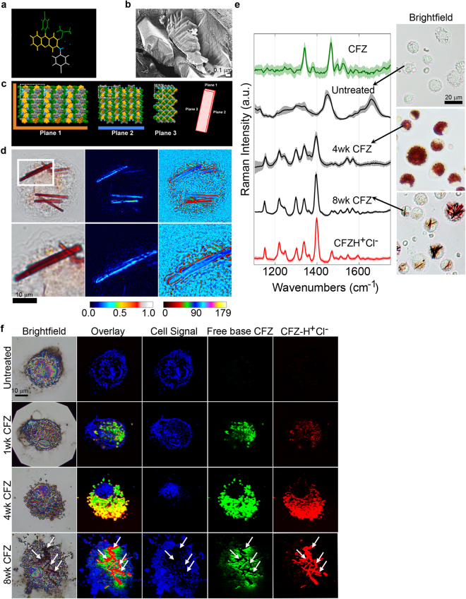
Figure 2.
Intracellular self-assembled crystal organization of CFZ-H+Cl−. (a) CFZ-H+Cl− chemical structure with color coding corresponding to the regions of the structure in the crystal arrangement. (b) Freeze-fracture electron micrograph of CLDIs within a Kupffer cell, showing that the CLDIs are sequestered inside the cell as multiple layered planes with lamellar spacing of 6 nm to 14 nm, by a double membrane of biological origin (~20 nm in thickness)21. (c) Crystal packing of CFZ-H+Cl− showing the three crystallographic planes of the crystal: a hydrophobic face in orange (plane 1), a hydrophilic face in blue (plane 2), and an amphipathic face (plane 3)8. (d) CLDIs (100–1.5X magnification) zoomed in (zoom factor of 2) in alveolar macrophages show high dichroism and an axis of highest transmittance, with a different profile (represented by the different signal intensity associated with the color bar) observed specifically around the edges of the crystal. (e) Raman spectra of free base CFZ, CFZ-H+Cl−, untreated and treated alveolar macrophages (4 and 8wks CFZ-fed mice) show distinct Raman peaks distinguishing between the different forms of the drug (red in the brightfield images): free base CFZ (1341 and 1465 cm−1), CFZ-H+Cl− (1399 cm−1). (f) Reflected brightfield and Raman images of alveolar macrophages from CFZ-fed mice prepared on silicon chip substrates; basis spectral fitting was used to show the temporal accumulation and cytoplasmic packaging of CFZ into highly ordered CLDIs.

