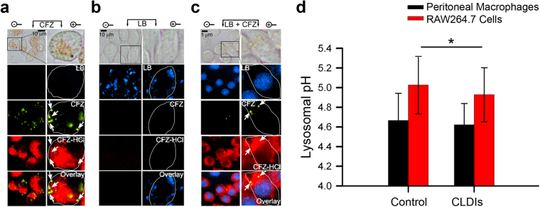
Figure 4.
Evidence of lysosomal accumulation of CFZ via fluorescence staining patterns in RAW264.7 cells. (a) Epifluorescence microscopy of RAW264.7 cells (40X magnification) zoomed in (zoom factor of 2.5) when incubated with (a) CFZ (10 μM), (b) Lysotracker Blue (LB, 1 μM), and (c) CFZ (10 μM) and LB (1 μM) together at t = 24 hours. Green fluorescence spots indicative of CFZ are also further annotated using white arrows for cross-referencing with other images. Note the lack of any far-red fluorescence positive signal in the direction pointed by the arrows. Vesicular staining pattern of LB visible as blue punctate spots in (b) are absent in (c), while nuclear staining becomes visible due to LB displacement from the lysosome to the nucleus because of CFZ accumulation in the lysosome. Cell boundaries are shown in white in the digitally zoomed panels. (d) Lysosomal pH measurements of RAW26.7 cells in the presence and absence of CLDIs, and peritoneal macrophages of control versus 8-week CFZ-fed mice. Data are mean ± S.D. of 29–181 measurements; *p = 0.026; analyzed by unpaired two-tailed Student t-test.

