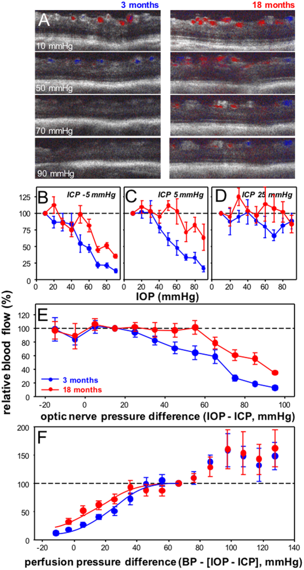
Figure 2.
Effect of IOP and ICP modification on inner retinal blood flow in 3 and 18 month old rats. (A) Representative Doppler blood flow for a young (3 month, left panels) and an older eye (18 months, right panels) to increasing IOP levels at an ICP of −5 mmHg. Group average (±SEM) normalized blood flow (against baseline IOP = 10 mmHg) for young and older eyes as a function of IOP at ICP −5 mmHg (B, 3 month n = 5, 18 month n = 5), ICP 5 mmHg (C, 3 month n = 5, 18 month n = 5) and ICP 25 mmHg (D, 3 month n = 5, 18 month n = 5). (E) Data for each eye is normalized against an ONPD of 15 mmHg. All data are combined for young and older eyes and plotted as a function of ONPD (mmHg). (F) Data for each eye is normalized against a perfusion pressure difference of 70 mmHg. All data are combined for young and older eyes and plotted as a function of optic nerve perfusion pressure difference (BP − [IOP − ICP], mmHg). Lines indicate dose response curve fits to 3 and 18 month old data.

