Figure 3.
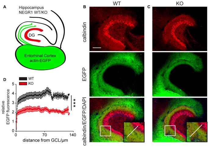
Entorhinal axon growth is impaired in the Negr1-deficient hippocampus. (A) Scheme illustrating the organotypic co-culture assay. (B,C) Anti-EGFP/anti-calbindin/DAPI co-labeling shows ingrowth of EGFP-expressing entorhinal axons into the DG of Negr1-wildtype (WT) mice (B) or Negr1-knockout (KO) mice. (B,C, insert) Images were processed for line scan analysis across the granule cell layer (GCL) with respect to calbindin-immunostaining as depicted. (D) Line scan analysis of GCL from WT (n = 41) and KO mice (n = 37) shows a decreased and less gradual entorhinal axon ingrowth in Negr1-KO mice. ***p < 0.001, Mann Whitney U test. Scale: 100 μm (B).

