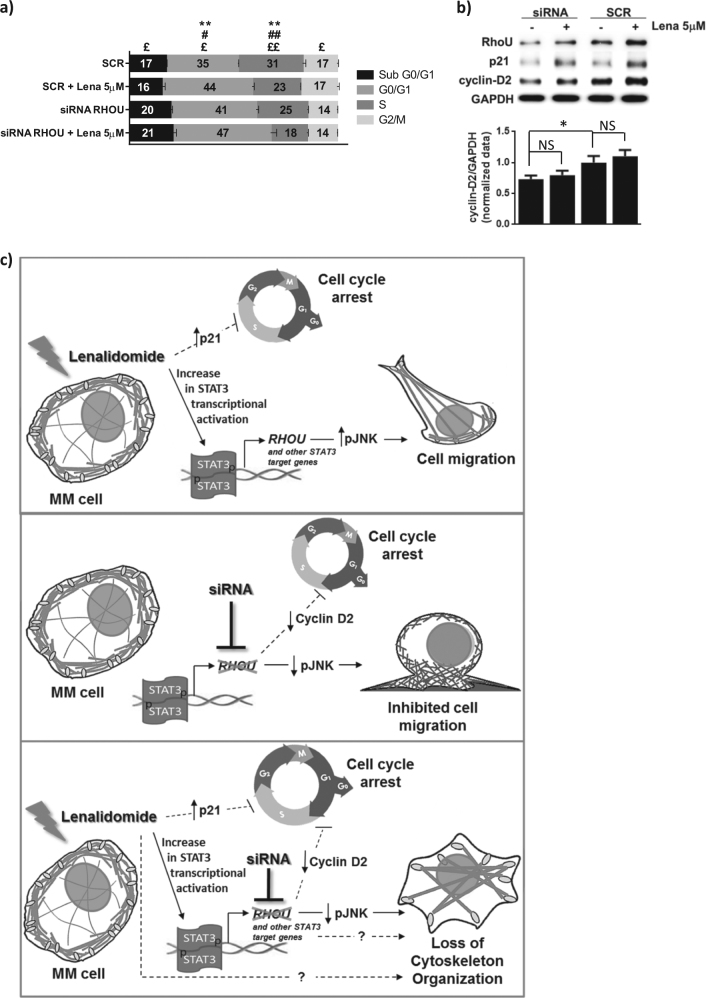
Fig. 6. Cell cycle analysis and schematic view of the effect of lenalidomide, RhoU silencing or their combination in MM cells.
a Cell cycle analysis based on PI staining of SCR and siRNA RHOU cells 48 h after transfection with the addition of lenalidomide in the last 24 h. Data represents mean ± SD of three independent experiments. Student’s t test: *SCR compared to SCR + Lena; #siRNA RhoU compared to siRNA RhoU + Lena; £siRNA RhoU + Lena compared to SCR + Lena, *p < 0.05; **p < 0.01. b Representative immunoblots from six samples from at least two independent experiments of RhoU, cyclin D2, and p21 expression after siRNA transfection and lenalidomide treatments, and densitometry of the expression of cyclin D2/GAPDH. c Lenalidomide treatment determines an increase in STAT3 activation and consequently the increased expression of STAT3 target genes including RhoU. This, combined with increased levels of pJNK, results in a reorganization of the cytoskeleton and boosts cell migration. These effects are parallel to cell cycle arrest, through a mechanism that is not explored in this manuscript. RhoU inhibition, on the other hand, results in decreased levels of active JNK leading to decreased cell migration rates and increased lamellipodial protrusions. RhoU silencing also caused MM cell cycle arrest due to a decrease in the expression of cyclin D2. The combination of both RhoU silencing and lenalidomide treatment further inhibits cell cycle progression and overcomes lenalidomide induced migration by decreasing the levels of active JNK causing however a loss of cytoskeleton organization. This cytoskeleton organization loss may be due to the expression of other STAT3 target genes or due to lenalidomide targets not explored in this article

