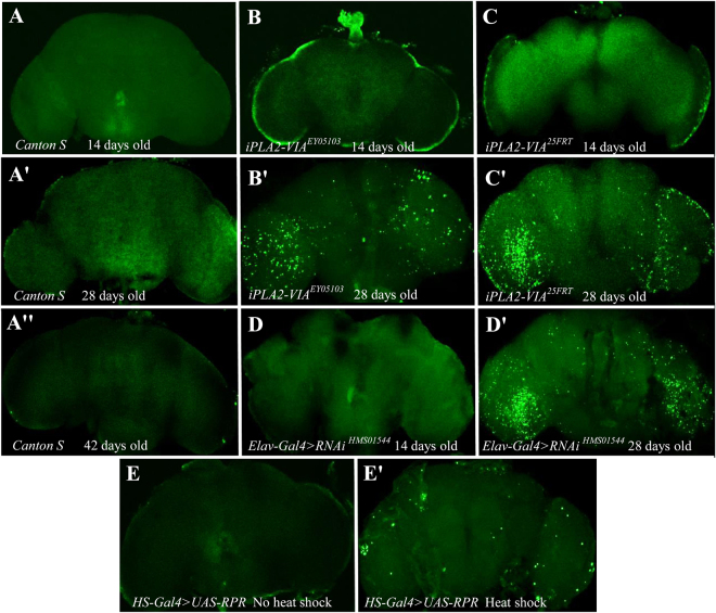
Figure 7.
iPLA2-VIA mutants show age-dependent neurodegeneration revealed by TUNEL staining. Confocal microscopy images of adult brains are shown. Canton S (A,A’ and A”) and UAS-rpr (E and E’) were used as negative and positive controls, respectively. UAS-rpr line was crossed with hs-GAL4 and raised at 18 °C. Their F1 progeny were also raised at 18 °C and 5–7 days after eclosion flies were heat-shocked at 38 °C for 20 min. 5 days after heat shock brains of heat-shocked (E’) and non-heat-shocked control flies (E) were analyzed. No neurodegeneration was found in the brains of 14 day old mutant (B and C) and pan-neuronal knockdown flies (D). Brains dissected from both 28 day old iPLA2-VIA25FRT null and P element insertional iPLA2- VIAEY05103 mutants as well as from 28 day old elav-GAL4 > RNAiHMS01544 knockdown flies (D’) show a very prominent number of apoptotic positive cells compared to physiologically and age- matched wild type Canton S controls (A,A’ and A”).

