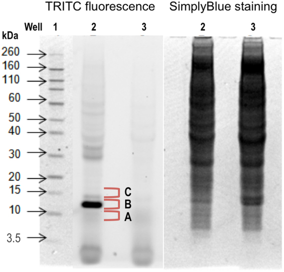
Figure 3.

Gel separation of proteins from lymph node cells. Lymp node cell proteins were isolated from pooled lymph node cells from mice exposed to vehicle (acetone:dibutyl phthalate, 1:1) or TRITC (5.6 mM) on the dorsum of the ears for three consecutive days. Proteins were separated using 1D SDS-PAGE and TRITC fluorescence (panel 1) was visualized using a fluorescence scanner with a GreenLED 528 nm laser with a bandpass filter of 605 ± 50 nm. Each well was loaded with 100 µg of lymph node protein from TRITC (well 2) or vehicle (well 3) exposed mice. The Novex Sharp pre-stained standard was used as a molecular standard (well 1). The total protein contents in wells 2 and 3 were visualized with SimplyBlue staining (panel 2).
