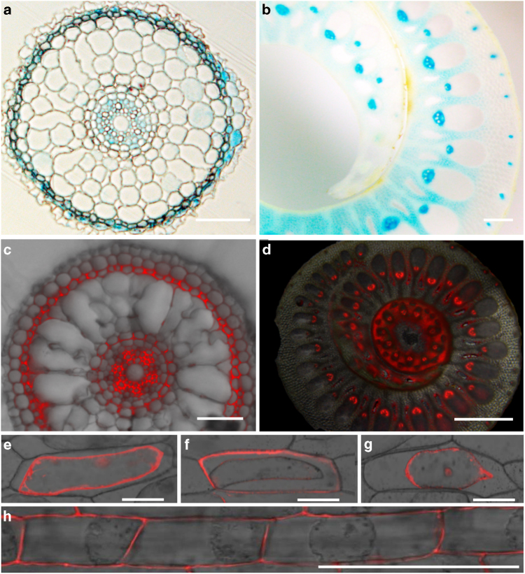
Fig. 3.
Histochemical and subcellular localization assay. a, b GUS expression driven by the CAL1TN1 promoter. (a) Cross section of 1 week old seedling roots. b Cross section of flag leaf sheath. c, d Fluorescence images of cross-sectioned seedling roots (c) and flag leaf sheath (d) from plants expressing the construct proCAL1TN1::CAL1-mRFP. e–g Subcellular localization of CAL1. Onion epidermal cells transiently transformed with mRFP (e), CAL1-mRFP fusion (f), or mRFP-CAL1 (g) were incubated in 40% sucrose to induce plasmolysis and then imaged by confocal microscopy. h Subcellular localization of CAL1 in rice plants harboring the construct 35S::CAL1-mRFP. Root epidermal cells were incubated in 40% sucrose to induce plasmolysis and then imaged by confocal microscopy. Bars = 1 mm in (b, g), 100 µm in (a, c–e, f, h)

