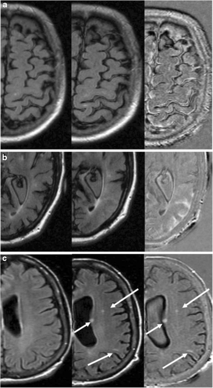
Fig. 2.
Details of three cases, with axial baseline FLAIR scan (left), the follow-up FLAIR scan (center) showing doubtful amyloid-related image abnormalities with vasogenic oedema and/or sulcal effusion (ARIA-E) findings and subtraction images (right) aiding in their detection (a), evaluation of their extent (b), and differential diagnosis (c). (a) Image artifacts prevent ARIA-E detection in FLAIR but the abnormalities are more visible on subtraction images. (b) The extension of the gyral swelling in the left occipital lobe is cumbersome to evaluate on FLAIR images but definitely more clear-cut in the subtraction images. (c) Small signal hyperintensities are visible on both FLAIR and subtraction images, hence their vascular origin may be excluded

