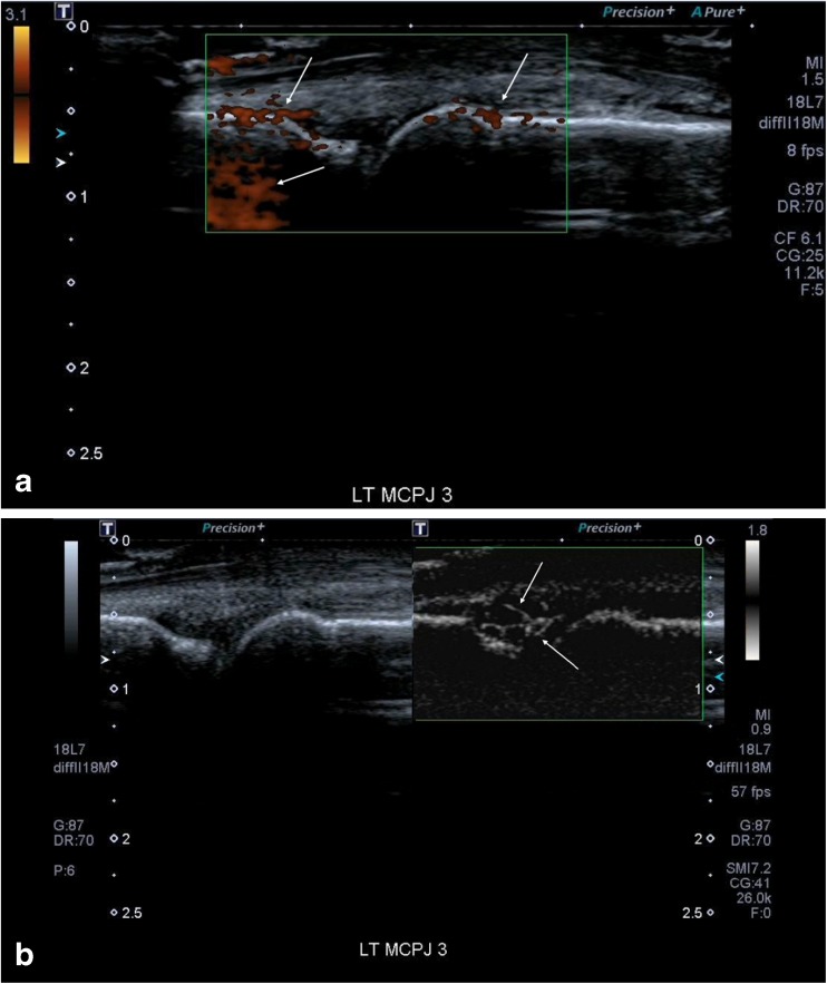
Fig. 2.
(a) This shows an image of a metacarpophalangeal joint (MCPJ) in a patient with an inflammatory arthritis and a symptomatic joint. Even with the Power Doppler gain turned right up and a low scale, resulting in much noise (white arrows), there remains no vascular signal within the joint to support an active synovitis. (b) There is much vascularity seen with Superb Microvascular Imaging (SMI) within the joint with fine spatial resolution (arrows). This would therefore denote an active synovitis in keeping with the patient’s symptoms and also elevated erythrocyte sedimentation rate (ESR) and C-reactive protein (CRP)

