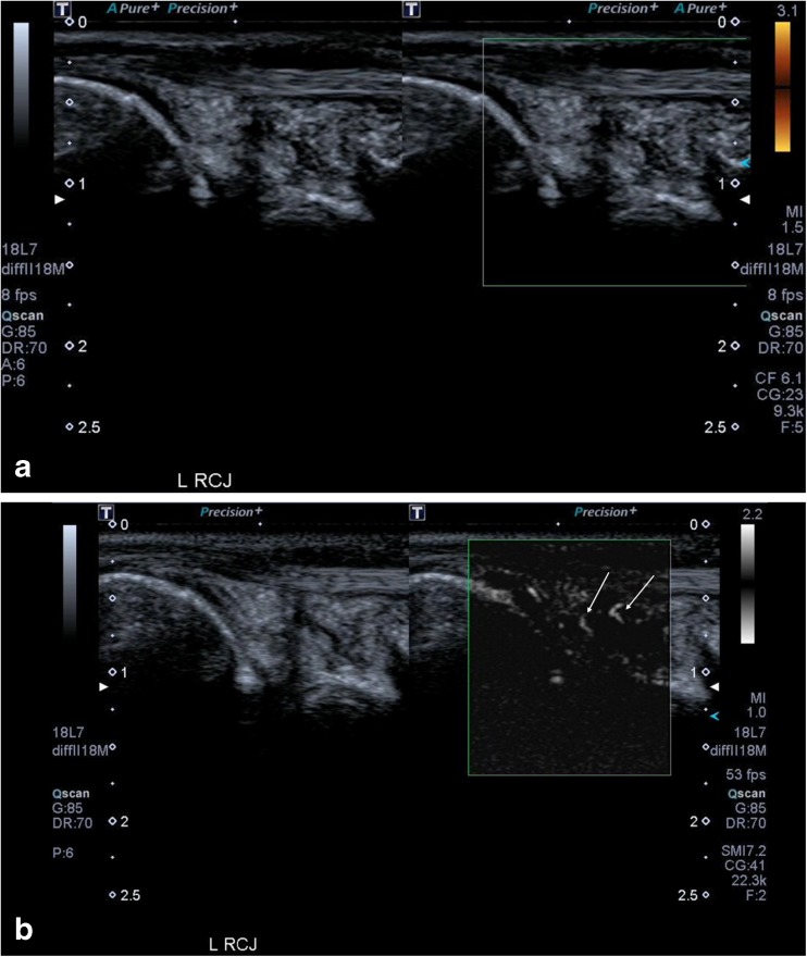
Fig. 3.
(a) These images show synovial hypertrophy on the grey-scale image of the radiocarpal joint (RCJ) in a patient with rheumatoid arthritis. In the dual image the right side of the split screen shows no detectable vascular flow within the thickened synovium on Power Doppler ultrasound (PDUS). (b) This second pair of images shows that there is clear neovascularity within the joint (arrows) on Superb Microvascular Imaging (SMI) indicating active inflammation, which was not evident on PDUS. The increased sensitivity and spatial resolution of SMI is much better appreciated on videoclips rather than still images and these have been included as supplementary material for this patient

