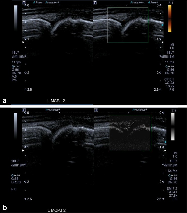
Fig. 4.
(a) These images show a relatively normal appearing joint on the grey-scale image of the metacarpophalangeal joint (MCPJ) of the left index finger, which was tender in this patient with rheumatoid arthritis. There is also no signal detected on Power Doppler ultrasound (PDUS). (b) The corresponding paired Superb Microvascular Imaging (SMI) images show that there is clear neovascularity seen within the joint (arrows) indicating active inflammation. This image highlights the resolution and flow within very small vessels that can be detected with SMI

