Figure 2.
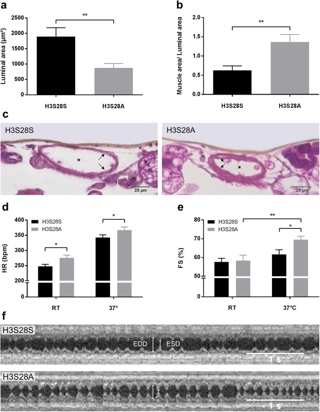
Histomorphological and functional analysis of H3S28S and H3S28A heart tubes. Quantification of the (a) luminal area and (b) heart muscle area over the entire cross section normalized to the luminal area (nH3S28S, H3S28A = 11, 7 days old). (c) Representative cross sections of H3S28S and H3S28A heart tubes at the level of the conical chamber. Arrows mark the heart wall; asterisks mark the heart lumen. (d) Heart rate and (e) fractional shortening (FS) as a measure for the cardiac contractile function. All measurements were performed at room temperature (RT) and following thermal stimulation (37 °C) for at least five minutes (nH3S28S = 22, nH3S28A = 24; 2 days old). (f) Exemplary M-mode OCT registrations from H3S28S and H3S28A flies following thermal stimulation. EDD: end-diastolic diameter; ESD: end-systolic diameter.

