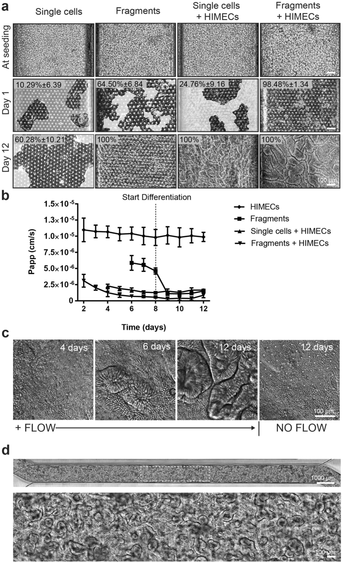
Figure 3.
Establishment of the primary human intestinal epithelium in the Organ Chip in the presence or absence intestine-specific microvascular endothelium. (a) Comparison of the efficiency of intestinal epithelial cell monolayer formation and development of villi-like structures between Organ Chip devices seeded with single intestinal epithelial cells or organoid fragments in the presence or absence of human intestinal microvascular endothelial cells (HIMECs). All chips were maintained for 1 or 12 days under continuous apical fluid flow of expansion (day 1–8) and differentiation media (day 9–12) and basal perfusion of EGM2-MV medium. White areas delineate empty spaces where initially plated cells detached from the membrane when flow was initiated; dark areas represent patches of attached cells. Graph shows the percentage of the area covered by epithelium at days 1 and 12 of culture, as assessed across 3 different field of views/chip with at least 3 chip replicates per condition, and expressed as mean ±SEM from 3 independent experiments. Note that when EGM2-MV medium was perfused basally, epithelium formed intestinal villi-like structures only when co-cultured with HIMECs seeded on the opposite side of the porous membrane. (b) Graph showing maturation of the intestinal barrier function on chips cultured under the same conditions measured by quantifying permeability of fluorescent Lucifer Yellow. Papp values are presented as mean ± SEM from 3 independent experiments; only Papp values for the cultures that reached 100% of confluency are plotted here. Note that the epithelium develops a higher barrier resistance to Lucifer Yellow more quickly when HIMECs are present. (c) Representative differential interference contrast images of the primary intestinal epithelium cultured on chip with HIMECs for 4, 6 or 12 days under continuous flow compared to 12 days in the absence of flow (note that formation of intestinal vill-like structures occurred only in the presence of flow). (d) Phase contrast microscopy views of the entire length of the epithelial microchannel (top) and a higher magnification of the area highlighted in the white rectangle (bottom) showing the human primary intestinal epithelium co-cultured with intestinal microvascular endothelial cells in the Organ Chip under peristalsis-like motions and flow for 12 days (culture medium was switched from EM to DM on day 8). Note the presence of villi-like structures across the entire length of the channel.

