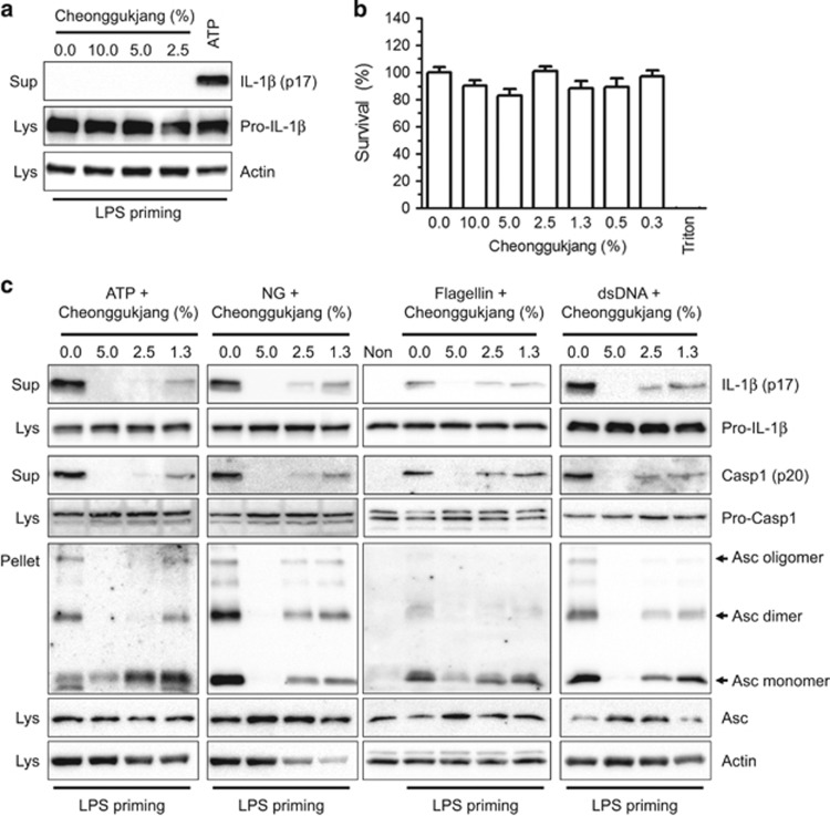
Figure 1.
Effect of cheonggukjang on inflammasome activation. BMDMs were primed with LPS (1 μg/ml) in RPMI medium containing 10% FBS and antibiotics for 3 h. (a) Cells were treated with medium containing the indicated percentages of cheonggukjang extracts or ATP (2 mm) and incubated for 1 h. The immunoblot assays were analyzed as indicated. (b) Cytotoxicity was measured after applying the indicated cheonggukjang dosage to BMDMs for 1 h, which was similar to the inflammasome activation step. Triton (0.01% of Triton x-100) was used to obtain complete cell death (0% survival rate), whereas the non-treated group was set as 100%. The data represent the mean±s.d. of three independent experiments, each performed in triplicate. (c) LPS-primed BMDMs were treated with ATP (2 mm), NG (40 μm), flagellin (0.5 mg/ml) or dsDNA (1 μg/ml) for 1 h in the presence of the indicated cheonggukjang concentration. Cellular supernatant (Sup), lysate (Lys) and cross-linked pellets (Pellet) from whole-cell lysates were analyzed with the indicated anti-sera in an immunoblot assay. All immunoblot data shown are representative of at least two independent experiments. BMDMs, bone marrow-derived macrophages; FBS, fetal bovine serum; LPS, lipopolysaccharide; NG, nigericin.

