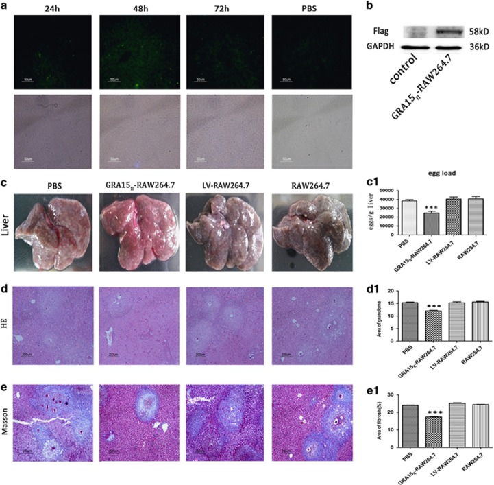
Figure 5.
GRA15II-RAW264.7 reached the liver tissues and survived. (a) Mouse liver frozen sections were observed under the fluorescence microscope. Samples were collected at 24, 48 and 72 h after GRA15II-RAW264.7 cell transfer. (b) The Flag tag was detected in mouse liver tissues 7 days after GRA15II-RAW264.7 cell transfer by western blotting. Pre-injection of GRA15II-RAW264.7 cells diminished liver pathological lesions in schistosome-infected mice. The livers were removed and shown in c. The egg load was lower in the GRA15II-RAW264.7-treated mice compared with the LV-RAW264.7-treated mice (c1). HE staining to show the granuloma area (d) and data analysis (d1). Masson staining was performed to assess the extent of hepatic fibrosis (e); the statistical analysis is shown in (e1). No significant difference in the pathological lesion index was observed between the LV-RAW264.7, RAW264.7 and PBS control mice. All asterisks indicate significant differences between the GRA15II-RAW264.7 and LV-RAW264.7-treated mice (***P<0.001, n=5). HE, hematoxylin and eosin; PBS, phophate-bufferred saline.

