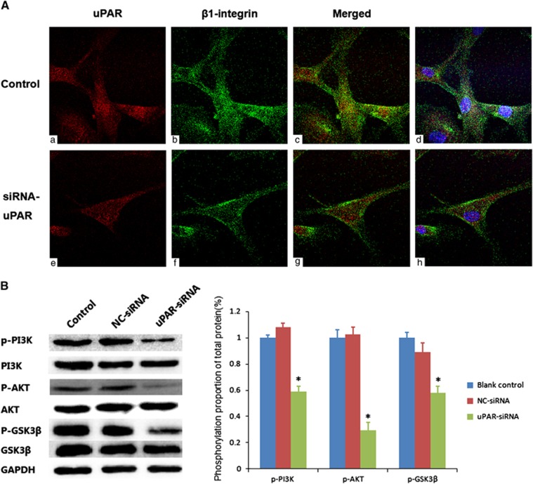
Figure 5.
(A) uPAR co-locates with β1-integrin and interferes its internalization. To facilitate visualization, images were converted to pseudo color using ImageJ software: uPAR (red, a, e), β1-integrin (green, b, f), and sites of colocalization (yellow, c, g). The nuclear staining was added in the merged images for easy location and shown on the right (blue, d, h). The above experiments were performed three times with similar results. (B) uPAR silencing reduces PI3K/Akt downstream signaling activation in RA-FLSs. Western blot showed the levels of phosphorylated PI3K, Akt and GSK3β in RA-FLSs after uPAR-siRNA (40 nM) or negative control (NC, 40 nM) following transfection for 72 h. Left panels show representative protein strips; right panels present densitometry quantification of protein expression (means±s.e.m.) of three independent experiments. *P<0.05, compared with the controls and n=4. RA-FLSs, fibroblast-like synoviocytes of rheumatoid arthritis; uPAR, Urokinase-type plasminogen activator receptor.

