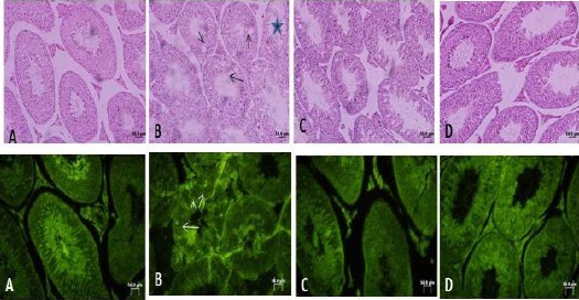
Figure 1.

Optical photomicrograph of mouse seminiferous tubules, A: Control, B: under treatment with ASA, C: under treatment with melatonin, D: under treatment with ASA+ melatonin. In the first row in control pay attention to active spermatogenesis in seminiferous tubules. ASA has caused a reduced quality of spermatogenesis. Black arrows in B show vacuoles inside the germinal epithelium. Large star shows a damaged tubule with thin epithelium in ASA treated mouse. In D note a better quality of seminiferous tubules. The first row is stained with Hematoxylin-Eosin, 200×. In the second row, white arrows show apoptotic bright yellow cells and a reduced level of bright yellow apoptotic cells in ASA+ melatonin-treated group compared with the ASA treated group. The second row is showing TUNEL staining, 400× Bar: 50 µm displayed improved morphology compared to the ASA-treated group. Sertoli cells and all types of germ cells were observed in the combined treatment group and vacuoles were not observed in the epithelial cells of seminiferous tubules from the combined treatment group (Figure 1). Combined administration of melatonin with ASA increased the quality and percentage of mature sperm and reduced the percentage of apoptotic cells compared to ASA treatment alone (P<0.001; Table 2).
