Figure 1.

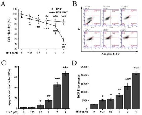
Effects of hypericin-photodynamic therapy on MH7A cell viability, apoptosis and production of intracellular reactive oxygen species. (A) Inhibition of cell viability by HYP-PDT was assayed using MTT. HYP-treated cells were subjected to PDT irradiation, and percent viable cells were normalized against untreated, non-irradiated cells (100%). Data are means±SD of n=5 independent experiments; *P<0.05, **P<0.01, ***P<0.001, compared to untreated, non-irradiated cells; #P<0.05, ##P<0.01, ###P<0.001, compared HYP-treated cells. (B, C) HYP-PDT induced apoptosis in MH7A cells. MH7A cells were treated with HYP (0–4 μM), and flowcytometry was used to measure apoptosis. HYP-PDT increased apoptosis and cell death in a concentration-dependent manner. Data are means±SD (n=3), *P<0.05, **P<0.01, ***P<0.001, compared with 0 μM HYP-PDT treated cells. (D) HYP-PDT enhanced ROS production in MH7A cells. Intracellular ROS production was measured using DCF 24 hr after treatment with increasing concentrations of HYP. DCF significantly increased with increasing HYP-PDT. With ≥1 μM HYP, DCF fluorescence increased and peaked with 4 μM HYP. Data were means ± SD (n = 3); *P<0.05, **P<0.01, ***P<0.001, compared to untreated cells (0 μM HYP)
DCF: dichlorofluorescein; HYP: hypericin; PDT: photodynamic therapy; ROS, reactive oxygen species
