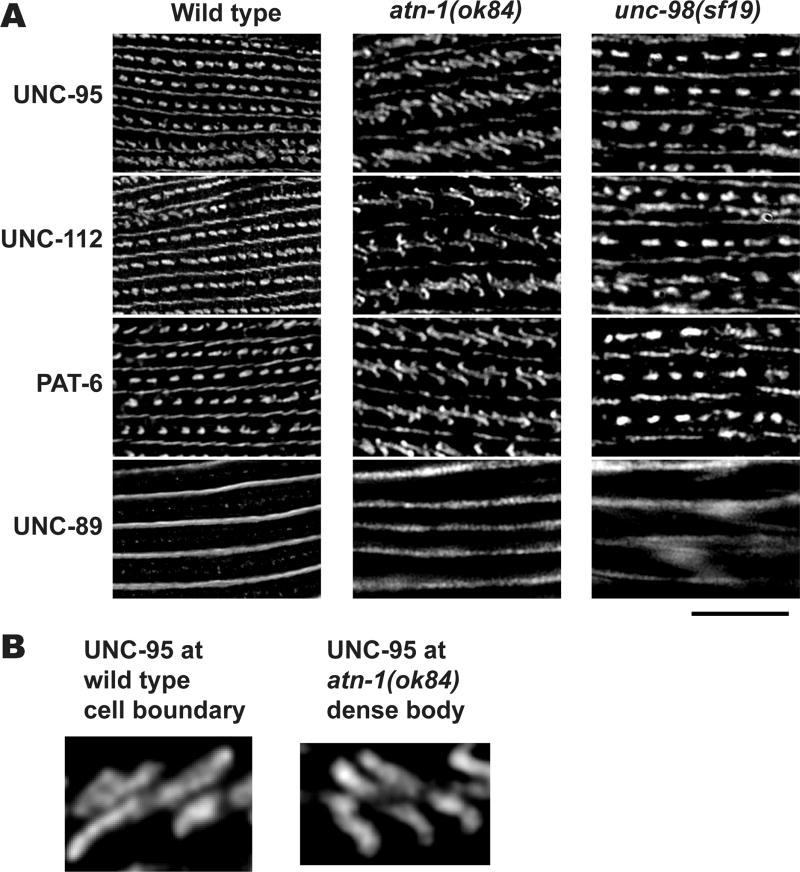
Figure 10. Lack of a deeper component of dense bodies changes their shape from “dot” like to “zipper” like.
(A) Wild type, atn-1 (ok84), and unc-98 (sf19) worms were immunostained with antibodies against UNC-95, UNC-112, PAT-6, and UNC-89. Anti-UNC-95, anti-UNC-112, and anti-PAT-6, localize to dense bodies and M-lines. Anti-UNC-89, localize only to M-lines. Images were captured by SIM. In the atn-1 (ok84) mutant, the dense bodies all appear like the “zipper” like structures normally found at muscle cell boundaries (compare with Figure 5). In contrast, in the unc-98(sf19) mutant, dense bodies mostly appear normal. Black scale bar represents 10 µm. (B) Enlargement of UNC-95 localization at a wild type cell boundary (left) and at an atn-1(ok84) dense body (right). Note the similarity in these two structures. Scale bar, 1 µm.

