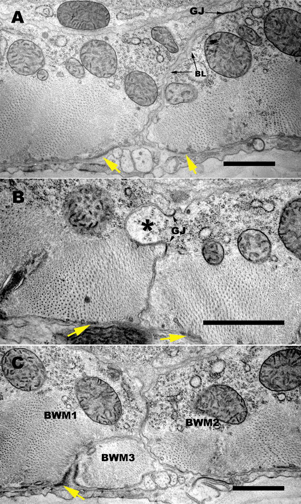
Figure 12. Several geometries along the lateral border contribute to the zipper-like structure there.
(A,B,C). Three transverse electron micrographs display some of the variety of features along the muscle cell-muscle cell apposition which help to produce a zig-zag pattern of the muscle protein localization observed by SIM. (A) Here the two adjoining muscle cells feature an overall slanting interface, from the region of the dense body at the basal surface to a large gap junction (GJ) near the apical limit of the border zone. Some basement membrane (BL) material fills the extracellular spaces here all the way to the gap junction. (B) Here a large “finger” of muscle tissue (asterisk) protrudes from one muscle cell into its neighbor, and two gap junctions (GJ) mark the base of this finger, connecting the two cells more intimately. (C) Here the border between two muscle cells (BWM1, BWM2) is invaded by the extreme distal portion of a third muscle cell (BWM3), so that the dense body material (marked with a yellow arrow) connects muscle cells 1 and 3, while the lateral border deviates sharply to allow muscle cells 1 and 2 to meet further in; note that BWM cells 1 and 2 are also connected by a gap junction (GJ). All of these geometries are common, and large enough to contribute to the flexible zig-zag shapes detectable by SIM. Scale bars, 1 µm.

