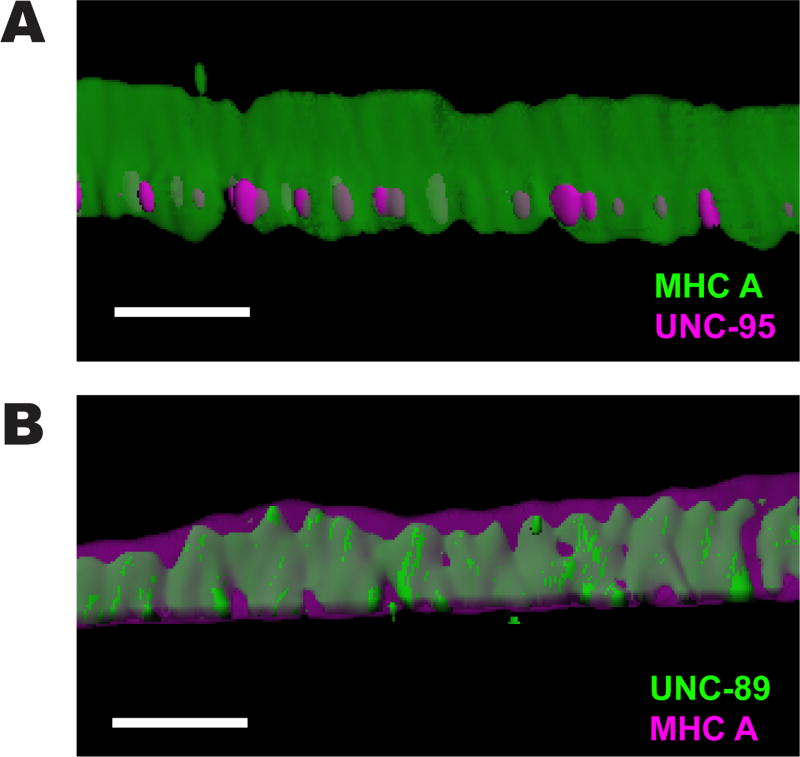
Figure 3. 3D rendering of localization of MHC A with UNC-95, and UNC-89 with MHC A.
(A) Side view of a single M-line immunostained with anti-MHC A (green) and anti-UNC-95 (magenta). MHC A also spans ~2 µm from near the muscle cell membrane deep into the sarcomere. Scale bar, 2 µm. (B) Side view of a single M-line immunostained with anti-UNC-89 (green) and anti-MHC A (magenta). Note that MHC A localizes beyond the localization of UNC-89. Scale bar, 2 µm.

